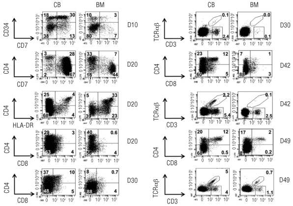
Figure 2.
Faster and more extensive T-cell differentiation by cord blood (CB) HSC. Sorted CD34+CD38−Lin− HSC from CB and bone marrow (BM) were cultured on OP9-DL1 cells in the presence of interleukin-7, Flt3 ligand and stem cell factor, and their developmental progression was assessed at the indicated time points by flow cytometric analysis for different antigens as indicated. Numbers in quadrants indicate the percentage of cells. TCR-αβ and TCR-γδ (CD3+-αβ cells) are delineated by regions, figures next to those regions indicate the corresponding percentage of cells.

