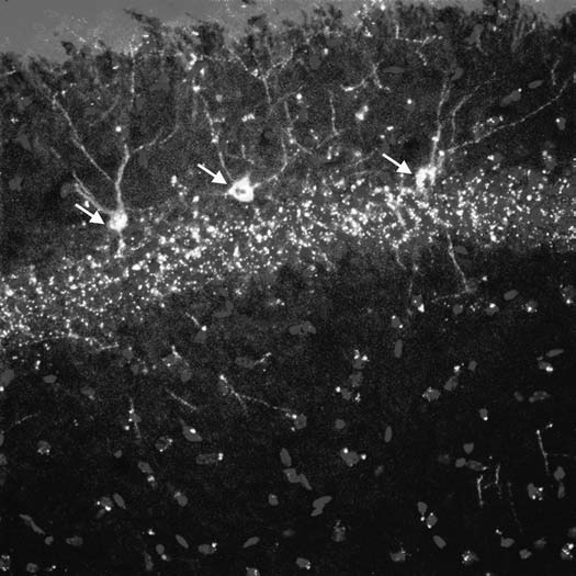
Figure 1.

Three PAPCs are shown (yellow, white arrows) coexpressing the fetal cell marker GFP and the neuronal marker β-3 tubulin in the cellular layer of the hippocampus of a murine mother. Note that PAPCs show axonal and dendritic aborizations, which are comparable to those from endogenous maternal hippocampal neurons. This indicates that PAPCs adopted a site specific identity.
