
Figure 10. Intermediate detection by pantetheine ejection ions.
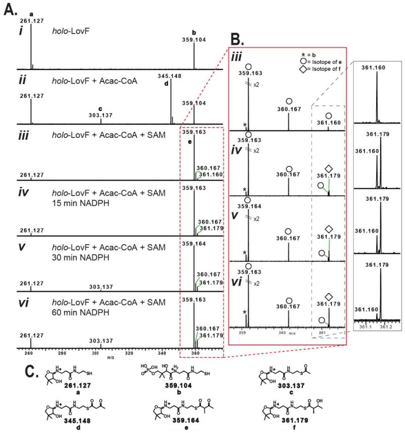
Comparison by FT-ICR-MS2 of various intermediate-loaded PPant ejection ions from holo-LovF following various in vitro incubation conditions. (A) PPant ejection ions resulting from specified incubation conditions. (B) Detail of ejection ions observed between 358-362 m/z by FT-ICR-MS. High resolution capabilities permit the differentiation of the α-methyl-acetoacetyl-loaded pantetheine ejection ion (e) and the phosphopantetheine ejection ion (b). Most critical is ability to detect the time-dependant formation and increase of low-abundance the β-hydroxy-α-methylbutyryl intermediate (361.179 m/z) and to distinguish this ion from an isotope 34S-containing isotope (361.160 m/z) of the α-methyl-acetoacetyl-loaded pantetheine ejection ion (e). An isotope of e containing a 13C was detectable at 360.167 m/z. (C) Detailed structures of various PPant ejection ions observed in the above spectra.