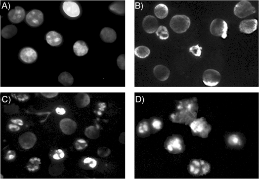
Figure 4.

Nuclear damage in KB-3-1 cells by treatment with 1 μm 12. Photos were taken by fluorescence microscopy after nuclei staining with Hoechst 33258. The figures show the microscopic morphology of the cells incubated in the presence of A) DMSO (0.1 % (v/v), control), and in the presence of 12 for B) 6, C) 18, and D) 24 h.
