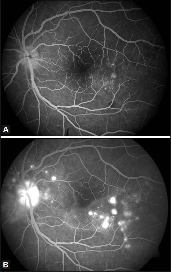
Figure 2.

(A) Fundus fluorescein angiography of the left eye showing multiple pinpoint hyperfluorescence in the early arteriovenous phase. (B) Fundus fluorescein angiography of the left eye showing pooling of dye in the subretinal space in the late arteriovenous phase
