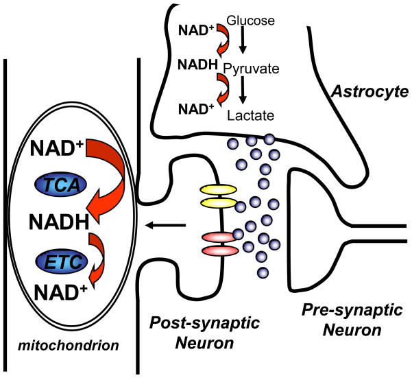
Figure 3.
Model to explain predominant NAD(P)H signals in neurons, rather than astrocytes, during synaptic signaling. It is likely that glutamate release triggers significant metabolic activation in both postsynaptic neurons and astrocytes. Glutamate activation of neuronal ionotropic receptors leads to significant mitochondrial activation, involving initial NADH decreases and subsequent overshooting NADH fluorescence increases (see Figure 2). These NADH dynamics in neurons are readily detectable, in part due to enhanced fluorescence of NADH in the mitochondrial compartment. Astrocyte glycolysis is likely activated following glutamate uptake, but any NADH increases from glycolysis in the cytosolic compartment appear difficult to detect, perhaps due to 1) small amplitude of fluorescence increases generated by NAD+/NADH transitions in the cytosolic compartment (see section 2) and/or 2) concomitant decreases in cytosolic NADH due to lactate generation.

