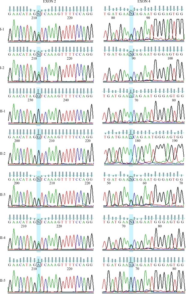
Figure 2.
Description of the mutations in the BCHE gene of the patient and her relatives. The figure shows the sequencing electropherograms of the amplified fragment of exons 2 and 4 of the analyzed individuals. The highlighted bases (blue) indicate the position of the mutations. The left column shows the results of the analysis of exon 2. Missense mutation GAT to GGT in codon 70 was found in heterozygosis in subjects I-1, I-2, II-3 and II-4, and in homozygosis in subjects II-1 and II-2 (patient). The right column shows the results of the analysis of exon 4. Missense mutation GCA to ACA in codon 539 was found in heterozygosis in individuals I-1, I-2, II-3 and II-4, and in homozygosis in individuals II-1 and II-2 (patient). Subject II-5 did not present mutations in any of both exons. Individuals are identified as in Figure 1.

