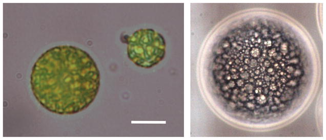
Fig. 2.

Micrographs of a W1/PFC/W2 emulsion containing fluorescein in the W1 phase. The left image is an overlay of both visible and fluorescent micrographs. The scale bar is 8 μm. The structure of the W1/PFC/W2 emulsion – water droplets containing fluorescein within a globule of PFC - can be clearly seen in the right image, which displays a 100 μm diameter globule.
