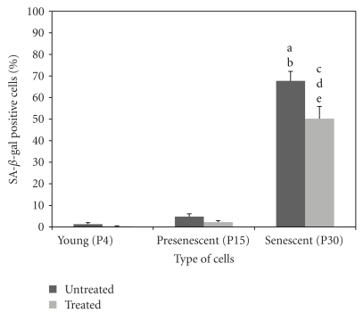
Figure 4.
Quantitative analysis of positive β-galactosidase stained cells in HDFs during cellular ageing. The percentage of cells positive for SA-β-gal staining was significantly increased in senescent cells. Incubation of senescent cells with TRF significantly decreased the percentage of cells positive for SA-β-gal staining. aDenotes P < .05 compared to untreated young HDFs, and bP < .05 compared to untreated presenescent HDFs, cP < .05 compared to untreated senescent HDFs, dP < .05 compared to treated young HDFs, eP < .05 compared to treated presenescent HDFs. Data are presented as mean ± SEM, n = 6.

