
Figure 5. Freshwater flagellate Cryptomonas.
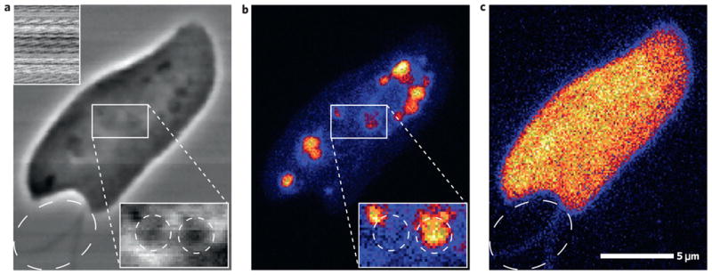
a, Scanning Zernike with absorption as inset. b,c, X-ray fluorescence of phosphorus (b) and sulphur (c). Magnified subregions in a and b are scaled separately from the main image to highlight features. The flagella (whiplike appendages) outside the cell are pointed out by a dashed ellipse in a and c.