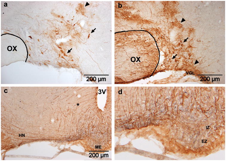
Figure 1. Adenoviral transduction in the SON.
GFP expression four days after SON injection of Ad-GFP. Two titers were tested: (a) 6.7×10⁷ pfu in 0.6μl or (b,c) 1.1×10⁹ pfu in 1.0μl. In both cases, transduced neurons (arrows) and glia (arrowheads) are present. Astrocytes in the VGL below the SON are also transduced (b). Stained fibers in the HN (c) and internal zone of the ME (d) confirm the presence of transduced SON MNCs. Stained tanycytes and their fibers (asterisk) are also visible (c). OX, optic chiasm; 3V, third ventricle; VGL, ventral glial lamina; ME, median eminence; HN, hypthalamo-neurohypophyseal tract; IZ, internal zone; EZ, external zone.

