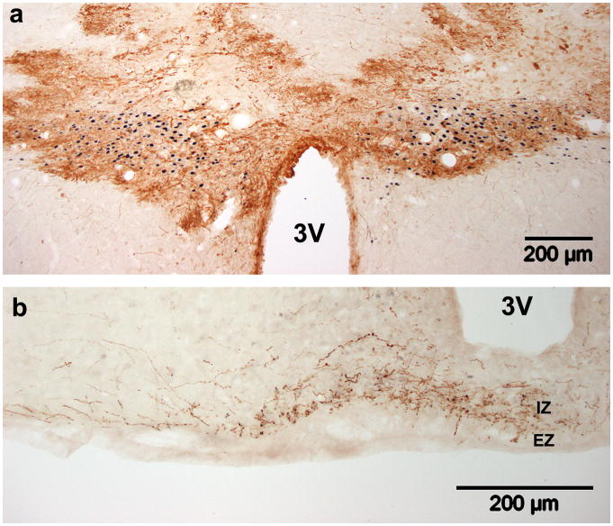
Figure 2. Adenovirus transduction in the PVN.
GFP expression seven days after bilateral injection of Ad-GFP (3.0×10⁶ pfu in 0.3μl) just above the PVN. (a) GFP expression (brown) in the PVN (delineated by black nuclear ERβ staining). (b) Though dense staining in the PVN prevents identification of affected cell types, fibers in the internal zone of the median eminence (ME) confirm transduction of MNC neurons, while fibers in the external zone indicate transduction of parvocellular neurons. 3V, third ventricle; IZ, internal zone; EZ, external zone.

