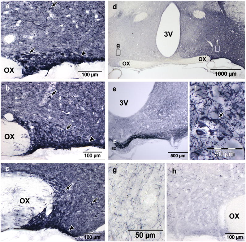
Figure 5. AAV2-CAG-GFP transduction in the SON four weeks after injection using convection enhanced delivery.
(a–c) GFP expression was very high in the anterior (a), mid (b) and posterior (c) SON after rapid injection (0.25μl/min) of 3.25×10⁹gp in 2.5μl. Arrows indicate neurons within or outside of the SON; arrowheads indicate the location of the ventral glial lamina. (d) A low power view of the hypothalamus shows that the dark staining is specific and restricted to the injected side. Fields of view for (f) and (g) are indicated. (e) Dense staining in the median eminence indicates efficient transduction of MNCs, though very dark staining in the SON is too dark to accurately count cells. Dark staining in the ipsilateral hypothalamus (a–d) is the result of a dense web GFP filled neurons and neurites (f). The contralateral hypothalamus also shows far fewer stained neurites and no cell bodies (g). Despite the large volume injected, the vector did not spread beyond the ipsilateral hypothalamus and, the contralateral hypothalamus (g) and SON (h) did not contain any GFP positive soma. OX, optic chiasm; 3V, third ventricle; IZ, internal zone; EZ, external zone.

