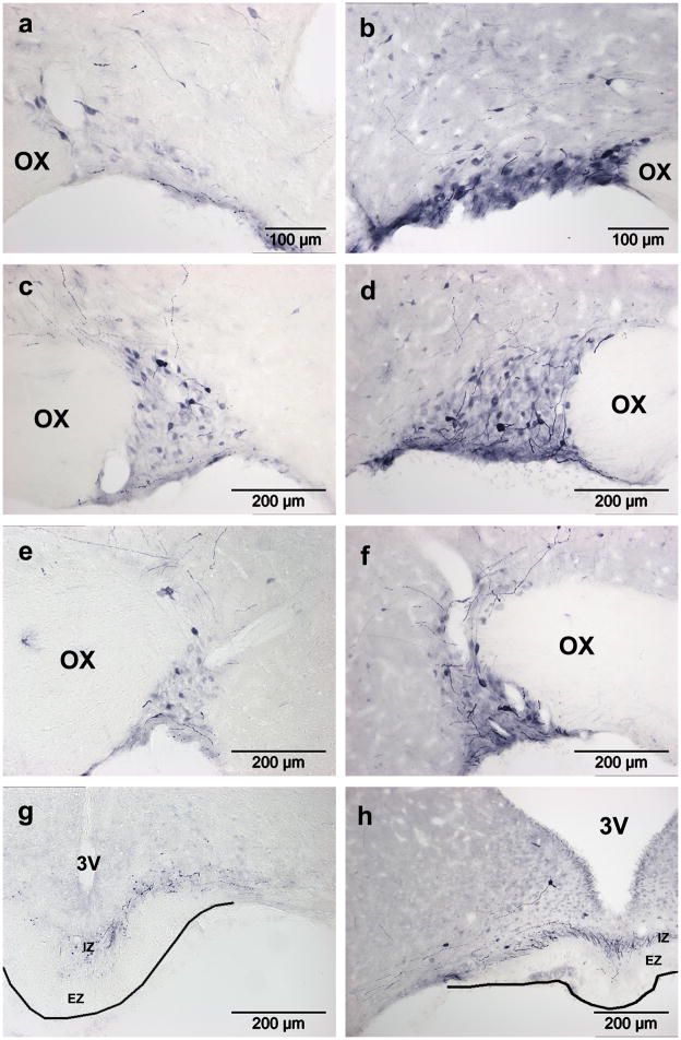
Figure 6. AAV5-GFP transduction in the SON after injection using convection enhanced delivery.
The number of GFP positive MNCs increased dramatically between three (a,c,e,g) and four (b,d,f,h) weeks post injection. At both timepoints, GFP positive MNCs were prominent in the anterior (a,b), mid (c,d) and posterior (e,f) SON, and there was no evidence of transduced glia. MNC axons are visible in the internal zone of the median eminence (g,h), and their sparseness is likely due to low GFP expression in neurites. OX, optic chiasm; 3V, third ventricle; IZ, internal zone; EZ, external zone.

