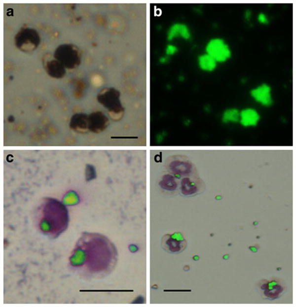
Fig. 1.

a Optical and b fluorescent microscopic image of monocytes labeled with MPIOs. These cells were labeled in culture following isolation from blood. Dual optical/fluorescent microscopic image of two monocytes (c) and five neutrophils (d) with internalized MPIOs. Some extracellular beads remain. Wright Giemsa-stained cytospun slides. All scale bars are 10 μm.
