
Fig. 2.
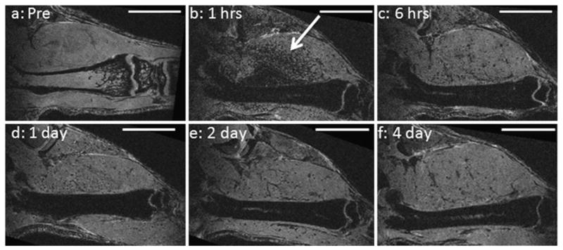
Single MRI slices extracted from 3-D data sets of same mouse femur a prior to injection of MPIOs, b 1 h, c 6 h, d 1 day, e 2 days, and f 4 days after injection of MPIOs. Bone marrow is dark from b–f. Bright streak in center of bone is blood vessel. Arrow points to dark, punctate contrast in muscle. All scale bars are 1 cm.