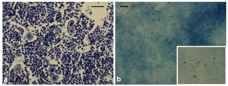
Fig. 4.

a Toluidine staining on decalcified tissue sections of bone marrow. b Unstained section where many small, brown particles are dispersed throughout the tissue. Inset is 3× magnification of parent image. Scale bars in a and b are 75 and 15 μm, respectively.
