Figure 3.
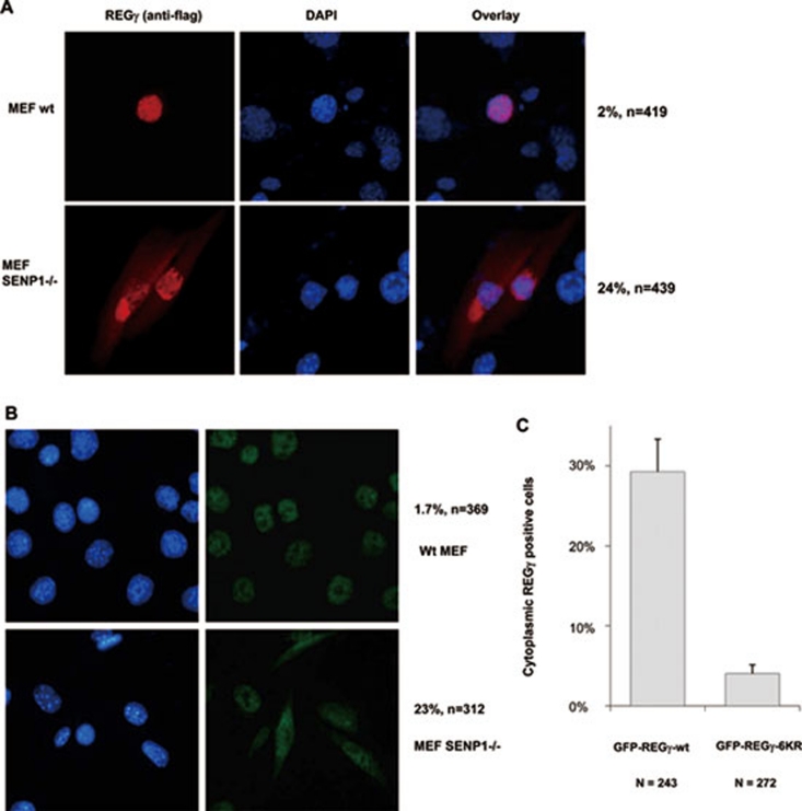
SUMO modification regulates cytosolic translocation of REGγ. (A) Cytosolic translocation of exogenous REGγ in SUMOylation-active cells. Wild-type and SENP-1−/− MEF cells were transiently transfected with pcDNA5/FRT/TO Flag-REGγ (1 μg). Twenty-four hours after transfection, immunostaining was performed with anti-DDK antibody (Flag) as described in Materials and methods to examine the expression and distribution of REGγ (red). DAPI staining indicates the location of nuclei. The percentage of transfected cells with cytoplasmic expression of REGγ is shown on the right. (B) SUMOylation enhances cytoplasmic translocation of endogenous REGγ. Wild-type and SENP-1−/− MEF cells were fixed and immunostaining was performed using anti-REGγ as in (A) to visualize cellular localization of endogenous REGγ. The percentage of REGγ-positive cells with cytoplasmic translocation is shown on the right. (C) SUMOylation-deficient REGγ has diminished cytoplasmic localization. SENP-1−/− MEF cells were transfected with 1 μg of GFP-REGγ or GFP-REGγ-6KR (mutation in all six predicated SUMO sites). The cytoplasmic vs nuclear fluorescent patterns in transfected cells expressing GFP fusion proteins were scored for statistical analysis. Data are shown as mean ± SD of three independent experiments (P < 0.01).

