
Figure 4.
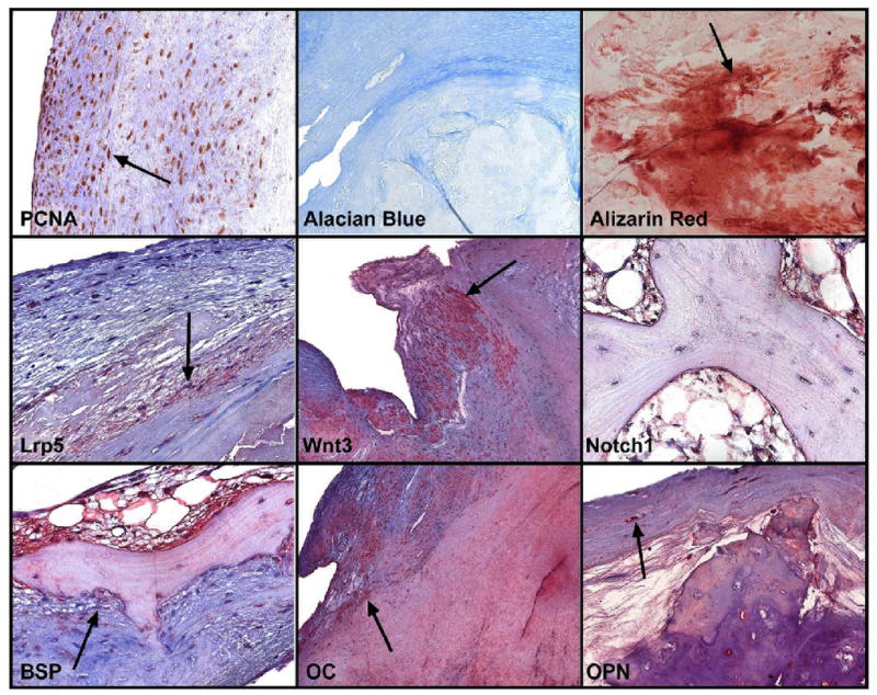
Bone immunohistochemistry for extracellular matrix and Lrp5/Notch1 signaling markers in Bicuspid Aortic Valves26. PCNA- Proliferating nuclear cell antigen indicates upregulation of DNA polymerase during active proliferation. Alcian blue stains for cartilage which is minimal. The stain binds to proteoglycans. The Alizarin red is a stain for calcium and hydroxyapatite in the valves which was increased markedly in the valves. The Lrp5 and Wnt3a stain is localized in the myofibroblast cells which are differentiating to an osteoblast like phenotype. The Notch1 stain is much less than the canonical Wnt markers in the areas of complex bone formation. The Bone sialoprotein, Osteocalcin and Osteopontin stains are all upregulated in bicuspid tissues indicating osteogenic differentiation and later stage mineralization. (Used with Permission from Elsevier, Publisher for Journal of the American College of Cardiology.)