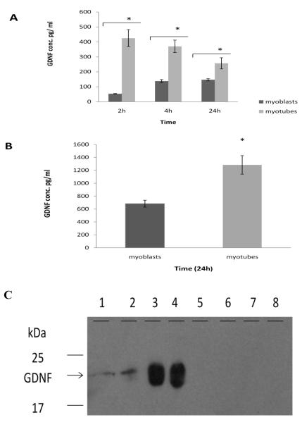
Figure 2. GDNF protein production in myoblasts, myotubes, and NG108-15 cells.
Samples of 3-day-old myoblasts or 10-day-old myotubes were taken at 2h, 4h, and 24h after changing medium. Myotubes were scraped from dishes at 2h, 4h, and 24h. Protein content in panel A and B was determined by ELISA. A. At all time points myotubes secreted significantly higher levels of GDNF than myoblasts. B. GDNF content within myoblasts and myotubes: Myotubes contain significantly more intracellular GDNF than myoblasts. Values are presented as mean ± S.E.M. Asterisk indicates significance (p≤ 0.05). n=4. C. Western blot: lanes 1& 2 represent GDNF secreted in culture medium by myotubes, lanes 3 & 4 represent GDNF contained within myotubes, lanes 5-6 represent GDNF in NG108-15 culture medium, and lane 7-6 represent GDNF in NG108-15 cells.

