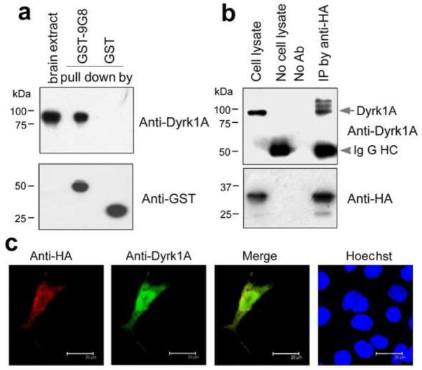
Fig. 5.
9G8 interacts with Dyrk1A. (a) Pull-down of Dyrk1A from rat brain extract by GST-9G8. GST-9G8 or GST coupled onto glutathione-Sepharose beads was incubated with rat brain extracts. After washing, the bound proteins were subjected to Western blots by using anti-GST and anti-Dyrk1A, respectively. (b) Co-immunoprecipitation of Dyrk1A with 9G8. HA-tagged 9G8 and Dyrk1A were co-expressed in COS7 cells for 48 h. The cell extract was incubated with anti-HA, and then protein G beads were added into the mixture. The bound proteins were subjected to Western blots by using antibodies indicated at the right of each blot. (c) Co-localization of 9G8 with Dyrk1A. HA-9G8 and Dyrk1A were co-transfected into COS7 cells. After 48 h transfection, the cells were fixed and immunostained by polyclonal anti-HA and monoclonal anti-Dyrk1A, followed by TRITC-anti-rabbit IgG (red) and FITC-anti-mouse IgG (green), respectively. Hoechst (blue) was used for nuclear staining.

