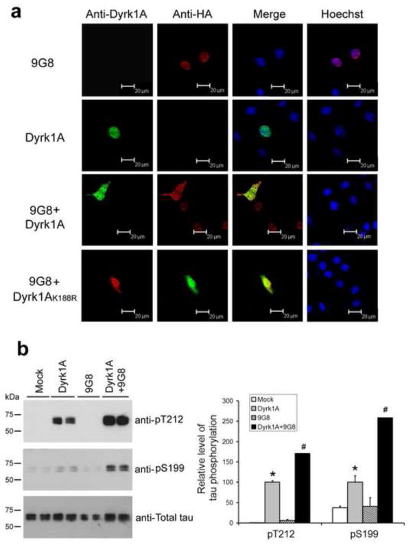
Fig. 7.
Co-expression of Dyrk1A or Dyrk1AK188R with 9G8 drives their translocalization from the nucleus into the cytoplasm. (a) COS7 cells were transfected, as indicated at the left of the figure. After 48 h transfection, the cells were fixed and immunostained by monoclonal anti-Dyrk1A and polyclonal anti-HA, followed by FITC-anti-mouse IgG (green) and TRITC-anti-rabbit IgG (red), respectively. Hoechst (blue) was used for nucleus staining. (b) HEK293FT cells were transfected with pCI-Neo/HA-Dyrk1A-Flag, pCEP4-9G8-HA and pCI-Neo/tau 40 for 48 h. The cells were then collected and the cell lysates were subjected to Western blots for measuring tau phosphorylation at T212 and S199. The blots were also quantified, and the data after being normalized with the total tau level are shown in graph at the right side. Data are presented as mean±SD (n=4). * P<0.05 versus mock control; # P<0.05 versus Dyrk1A alone.

