
Fig 2.
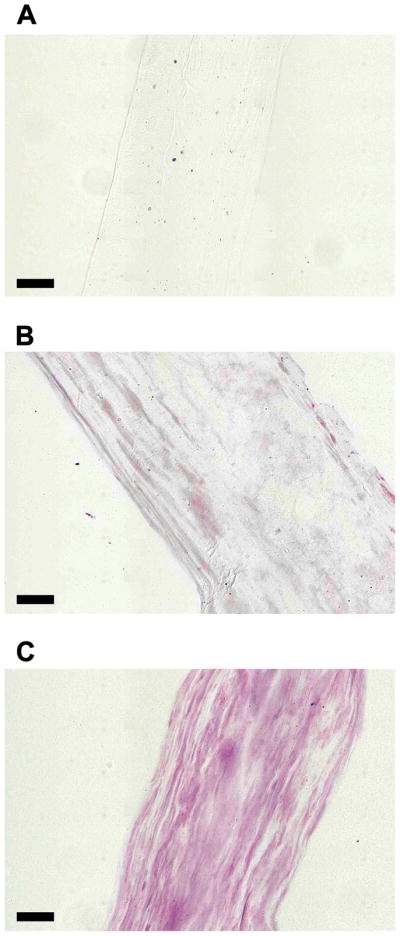
DMMB stained cryo-sections of ELAC. (A) Collagen Only (B) 30:1 Collagen:DS-SILY and (C) 10:1 Collagen:DS-SILY. DMMB staining revealed that DS-SILY was distributed throughout the length of the ELAC thread for both the 30:1 (B) and 10:1 (C) groups. The collagen only control (A) did not stain and was hardly visible under the microscope due to the transparency of the sample. (Scale bar: 50 μm).