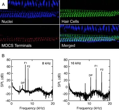
FIG. 9.
Normal structure and function of outer hair cells in a noise-reared α9KO mouse. A Middle turn of the cochlea showing a normal complement of outer hair cells and MOCS terminals. Hair cells were immunolabeled with antibodies for DAPI (blue) and myo7a (green). Efferent terminals were immunolabeled with SV2 (red). B Distortion product otoacoustic emissions produced by primary tones (F1 and F2) at frequencies near 8 and 16 kHz. A cubic difference distortion product (DP) was generated at both frequencies.

