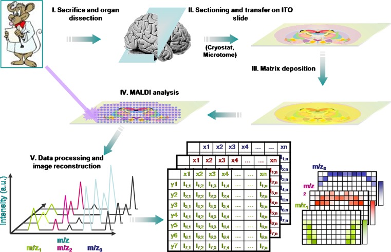
Fig. 3.
Overview of MALDI-MS imaging for tissue-based cryptomics. In MALDI-MS imaging, 10–20-μm-thick frozen tissue sections are cut using a microtome and transferred to a conductive surface. After matrix is deposited onto the section, MALDI ionization is carried out in the defined regions of the tissue section and spectral data acquired. The mass spectrometry data are computationally processed to generate a pseudoimage, showing abundances of individual peptides and proteins. a.u. arbitrary units, ITO iridium tin oxide (reprinted with permission from (31))

