
Fig. 1.
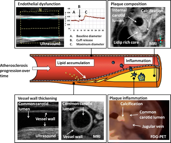
Various imaging modalities that can address different processes in atherosclerosis. Upper left: brachial artery endothelial function measurement with ultrasound (courtesy of Eric de Groot). The brachial artery diameter is measured at baseline and after cuff release and used to calculate the flow-dependent vasodilatation. Lower left: carotid vessel wall thickness measurement by ultrasound on the left (courtesy of Eric de Groot) and MRI on the right (courtesy of Aart Nederveen). Upper right: MRI of carotid artery plaque composition (courtesy of Aart Nederveen). Lower right: 18F-fluordeoxyglucose positron emission tomography (FDG-PET)/CT imaging of carotid artery plaque. Plaque calcification is visible on the CT scan (courtesy of Jan Bucerius)