
Fig. 8.
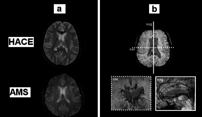
Susceptibility-weighted T2*-images (a) obtained in one subject 7 weeks after CT diagnosis of HACE following ascent to 3,860 m and shortly following clinical diagnosis of severe AMS in a separate subject at 5,895 m. Note the multiple patchy hypo-intensities (arrows) present in the HACE subject only that correspond to micro-hemorrhages that result in hemosiderin [insoluble Fe(III) oxide hydroxide] deposition secondary to blood–brain barrier disruption and cerebral capillary “stress failure”. These “micro-bleeds” were located predominantly in the genu and splenium of the corpus callosum (b) which have previously been identified as predilection sites for vasogenic edema in HACE [2]. Data modified from [69]