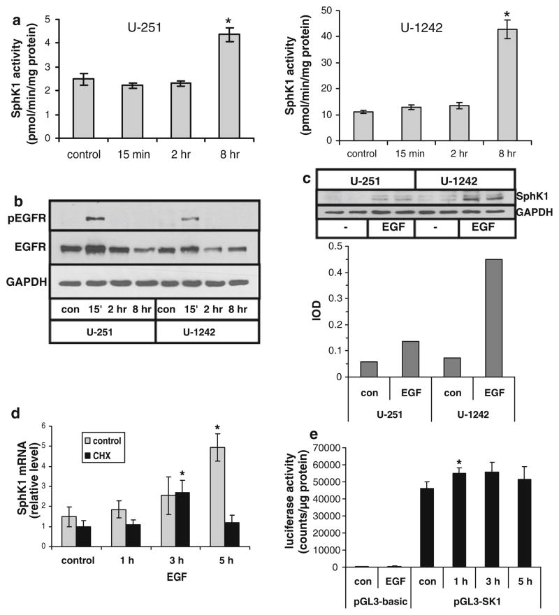
Fig. 1.
Regulation of SphK1 activity and expression by EGF in glioma cell lines. U-251 or U-1242 human glioma cells were stimulated with 20 ng/ml EGF for the indicated time. a SphK1 activity was measured as described in Materials and Methods. b Cell lysates were immunoblotted for EGFR and tyrosine phosphorylated EGFR. c Cells were treated for 8 h with or without 20 ng/ml EGF. Duplicate samples were immunoblotted for SphK1. Bands were quantitated by scanning densitometry, and results are ratios of IOD for SphK1 relative to GAPDH. d U-251 cells were pretreated for 30 min with 10 μg/ml cycloheximide or with ethanol as a vehicle control and then stimulated with EGF for the indicated time. RNA was isolated and SphK1 mRNA level was determined by real time PCR analysis. e U-251 cells were transfected with pGL3-basic or pGL3-SK1 and then replated in multiwell plates. Cells were stimulated with 20 ng/ml EGF for 3 h (pGL3-basic) or the indicated time (pGL3-SK1) and luciferase activity was measured. Results for panels a, d and e are means ± s.d. of triplicate samples. The * indicates statistically significant difference by Student’s T test, p < 0.05. Two independent experiments provided similar results for all panels

