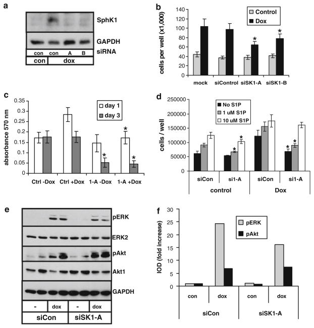
Fig. 3.
Role of SphK1 in EGFRvIII-mediated glioma cell proliferation. U-251-E18 cells were transfected with control random sequence siRNA (con) or two different siRNAs specific to SphK1 (A and B) and grown for 2 days in the absence or presence of 10 ng/ml dox. a Cell lysates were immunoblotted for SphK1. b Cell number was determined after 3 days in culture. c Cell proliferation was examined by MTT assay. d Cells were transfected with control or SphK1-specific siRNA and grown for 3 days in the absence or presence of dox and the indicated concentration of S1P. The * indicate statistically significant difference of SphK1-specific siRNA-transfected cells from control siRNA transfected cells, p < 0.05. e U-251-E18 cells were transfected with control siRNA or SphK1-specific siRNA and grown for 1 day in the absence or presence of dox. Duplicate cell lysates were immunoblotted for phosphorylated ERK or Akt, after which blots were stripped and reblotted for total ERK or Akt. f Western blots shown in panel d were quantitated by densitometry. Results are expressed as doxycycline-induced fold increase in the ratio of the integrated optical density (IOD) of phosphorylated to total ERK or Akt. At least two independent experiments provided similar results for all panels

