Fig. 3.

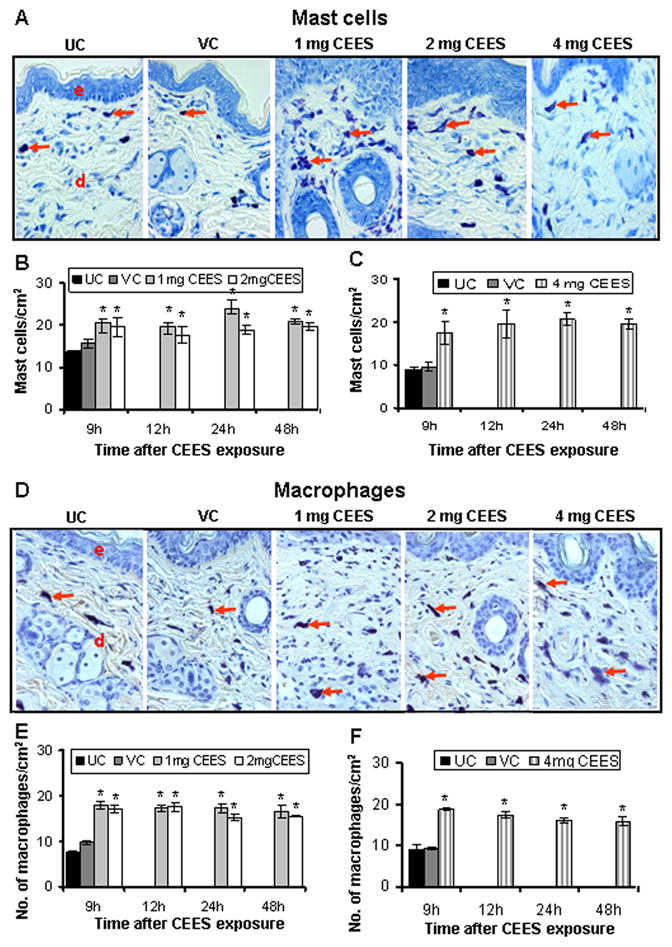
CEES topical exposure causes an increase in the number of mast cells and macrophages in male SKH-1 hairless mouse skin. Following 1, 2 or 4 mg CEES and control exposures, skin samples were collected as a function of time, and processed and stained as detailed under Materials and Methods. Representative toluidine blue stained mast cells (A) and F4/80 stained macrophages (D) in the skin sections from untreated and vehicle controls, and 1, 2 or 4 mg CEES exposed groups (×400 magnification). Mast cells/cm2 (B and C) and macrophages/cm2 (E and F) were calculated from various treatment groups by counting positive stained cells in randomly selected five fields per sample (×400 magnification) as detailed under Materials and Methods. Data presented are mean ± SEM of five animals of each group. Statistical significance of difference between the CEES exposed and control groups were determined by one way ANOVA followed by Bonferroni t-test for pair wise multiple comparisons. *, p<0.001; $, p<0.01 as compared to untreated control group. UC, untreated control; VC, vehicle control; e, epidermis; d, dermis; red arrows, toluidine blue or F4/80 positive cells.
