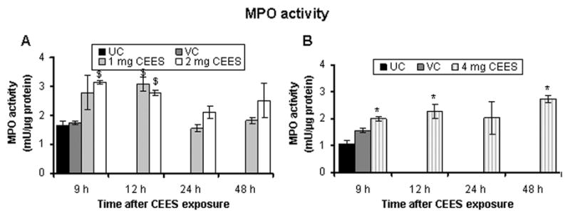
Fig. 4.

CEES topical exposure causes an increase in MPO activity, indicating neutrophil infiltration, in male SKH-1 hairless mouse skin. Following 1, 2 (A) or 4 (B) mg CEES and control exposures, skin samples were collected as a function of time, and MPO activity was determined using a fluorescence assay kit as described under Materials and Methods. Data presented are mean ± SEM of three-five animals in each treatment group and samples were taken in duplicate for the MPO assay. Statistical significance of difference between the CEES exposed and control groups were determined by one way ANOVA followed by Bonferroni t-test for pair wise multiple comparisons. *, p<0.001; $, p<0.01 as compared to untreated control group. UC, untreated control; VC, vehicle control.
