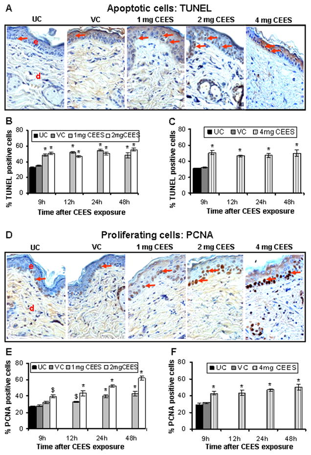
Fig. 5.

CEES topical exposure causes an increase in both apoptotic and proliferating cells in male SKH-1 hairless mouse skin. Following 1, 2 or 4 mg CEES and control exposures, skin samples were collected as a function of time, and processed and stained for apoptotic cell death (TUNEL) and cell proliferation (PCNA) as detailed in Materials and Methods. Representative TUNEL (A) and PCNA (D) stained skin sections from untreated and vehicle controls, and 1, 2 or 4 mg CEES exposed groups (×400 magnification). Percent TUNEL positive cells (B and C) and percent PCNA positive cells (E and F) were calculated from various treatment groups by counting positive stained cells in randomly selected five fields per sample (×400 magnification) as detailed in Materials and Methods. Data presented are mean ± SEM of five animals of each group. Statistical significance of difference between the CEES exposed and control groups were determined by one way ANOVA followed by Bonferroni t-test for pair wise multiple comparisons. *, p<0.001; $, p<0.01 as compared to untreated control group. UC, untreated control; VC, vehicle control; e, epidermis; d, dermis; red arrows, TUNEL or PCNA positive cells.
