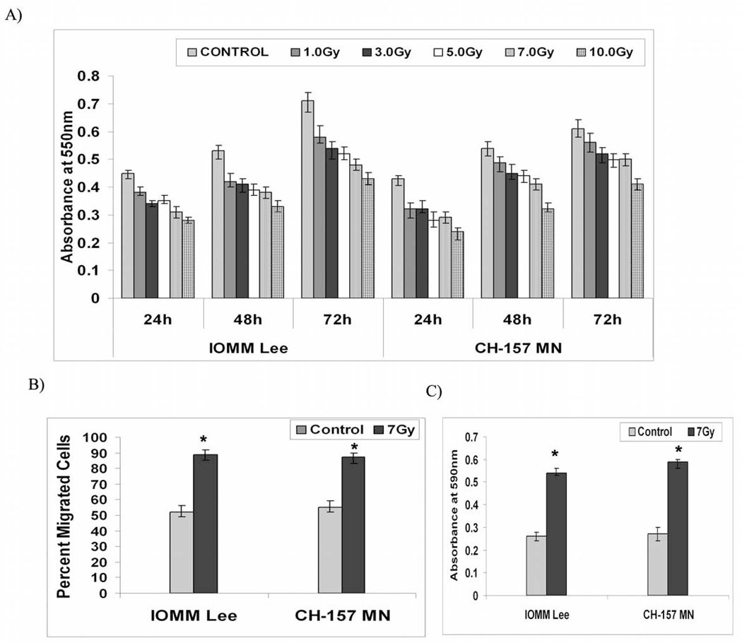
Figure 1. Radiation induces migration and decreases proliferation of meningioma cells.
A) IOMM-Lee and CH-157-MN cells (1×10⁵) were seeded in 6-well plates and irradiated at different doses of radiation as indicated. Then, cells were trypsinized and seeded at 1×10⁴ cells per well in 96-well plates. After the indicated hours of incubation, MTT reagent was added and followed by another 4 hrs of incubation and addition of acid-isopropanol. Absorbance was measured at 550 nm and the values were quantified. B) IOMM-Lee and CH-157-MN cells (1×10⁵) were seeded in 8-µm pore size transwell inserts, irradiated and allowed to migrate for 24 hrs. Later, cells that remained in the upper chamber and those that attached to the other side were fixed, stained and counted under a light microscope. The percentage of migrated cells was calculated. C) IOMM-Lee and CH-157-MN cells (2×10⁵) were seeded in 3-µm pore size transwell inserts, irradiated and allowed to migrate through the membrane for 24 hrs. Cells were then fixed and placed in crystal violet; cell bodies that remained on the top side of the membranes were scraped, the dye retained by the migration front on the lower side was extracted in 10% acetic acid, absorbance was measured at 590 nm, and the relative migration front was plotted. Values are mean ± S.D. from three independent experiments. * Statistically different compared to control and radiated groups (P<0.05)

