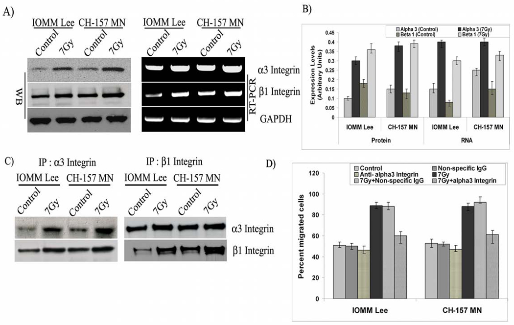
Figure 2. α3 and β1 integrins are overexpressed and involved in radiation-induced migration.
A) IOMM-Lee and CH-157-MN cells (1×10⁵) cells were seeded in 6-well plates, irradiated and incubated for 24 hrs. The cell lysates were subjected to western blot analysis using anti-α3, anti-β1 and GAPDH antibodies and quantified. Similarly, total RNA was extracted and subjected to RT-PCR for α3, β1, and GAPDH with specific primers. B). The expression levels of protein and RNA were quantified using image J software. C) IOMM-Lee and CH-157-MN cells (1×10⁵) were seeded in 8-µm pore size transwell inserts and treated with α3β1 antibody (10 µg/mL) one hour preceding the radiation treatment and allowed to migrate for 24 hrs. Cells that remained in the upper chamber and those that attached to the other side were fixed, stained and counted under a light microscope. The percent of migrated cells was calculated. D) IOMM-Lee and CH-157-MN cells (1×10⁵) cells were seeded in 6-well plates, irradiated and incubated for 24 hrs. The cell lysates were subjected to immunoprecipitation and followed by western blot analysis with the anti beta 1 and alpha 3 integrin antibodies. Values are mean ± S.D. from three independent experiments. * Statistically different (P<0.01)

