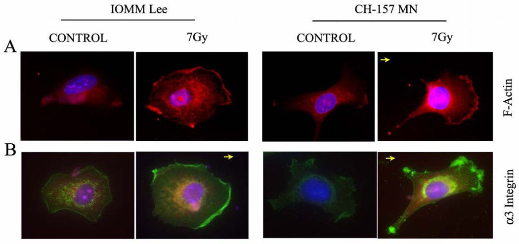
Figure 3. F-actin and α3 integrin localized to the leading edge in migrating cells.
Fluorescent micrograph of irradiated (7 Gy) IOMM-Lee and CH-157-MN cells that were grown for 24 hrs and stained with TRTc-labeled phalloidin and anti-α3 integrin antibody to mark localization. A) Pink fluorescence indicates where F-actin localized in the cell. B) Green fluorescence indicates the localization of α3 integrin, which is enriched at the leading front of migrating cells and in focal adhesions at the membrane edge of the extending lamellipodium. DAPI was used for nuclear staining. Images are representative pictures of several cells. Arrow indicates direction of cell movement.

