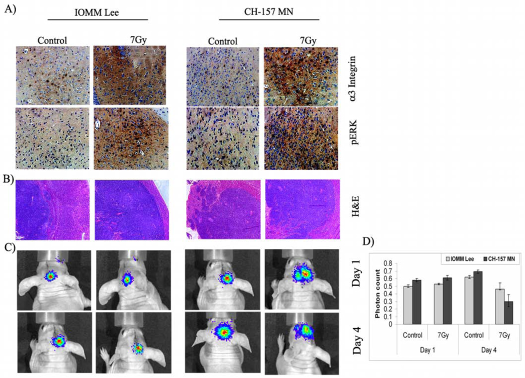
Figure 6. Migration in vivo.
IOMM-Lee and CH-157-MN cells were implanted intracranially in nude mice and treated with radiation as described in Materials and Methods. A total of 5 animals were studied in each group and 3 weeks after radiation treatment, animals were perfused and the brains were harvested and processed. A) Immunohistochemical analysis for α3 integrin and pERK in the paraffin-embedded sections of intracranial tumors implanted with IOMM-Lee and CH-157-MN cells. Slides were counterstained with hematoxylin for nuclear staining (40X). B) Tissue sections were stained with H&E as per standard protocol, and a representative tumor volume is shown (20X). C) IOMM-Lee and CH-157-MN luciferase-expressing stable cells (1×10⁵) were implanted into nude mice (4–6 weeks old). The first group was treated with cells irradiated at 7 Gy and the second group was infused with non-irradiated cells. Tumor progression was followed for one week with daily in vivo imaging. D) The luciferase activity from both groups of animals was quantified as photons per second. Values are mean ± S.D (n=5)

