
FIGURE 7.
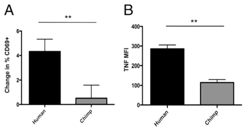
Exposure of PBMCs to ssRNA leads to more TNF production and increased cell surface expression of CD69 in human cells. Freshly isolated PBMCs were exposed to ssRNA for 20 h. A, Lymphocyte CD69 cell surface expression was analyzed by flow cytometry. Change in percent CD69 positive was determined by subtracting the percentage of CD69 positive cells in un-stimulated controls from the percentage of CD69 positive cells in stimulated samples. Data represent the mean of three experiments. Error bars represent SEM. B, Intracellular TNF expression was detected by flow cytometry. Data represent mean of three experiments. Error bars represent SEM. **p < 0.01.