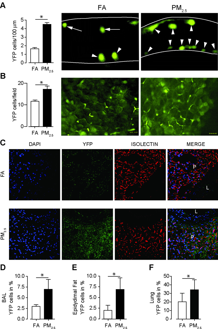
Figure 5.
Chronic PM2.5 exposure over 20 weeks increases monocyte adherence within microvasculature and tissue niches in c-fmsYFP mice (FVB/N background). (A) Representative images and quantification of adherent YFP cells in the cremasteric venular endothelium (open arrow heads adherent monocytes; arrow heads with tail, rolling monocytes). (Original magnification 400×, n=5). (B) Representative images and quantification of adherent YFP cells in the mesenteric adipose tissue. (Original magnification 200×, n=5). (C) Immunohistochemical staining for YFP positive monocyte infiltration into perivascular fat tissue in c-fmsYFP mice exposed to FA or PM2.5. Perivascular fat tissue from mice that express a yellow fluorescent protein (c-fmsYFP, yellow) was stained with DAPI (blue) and isolectin (red) and visualized by confocal microscopy. (L=lumen; P=perivascular fat). (Original magnification 400×, n=3). Quantification of YFP positive cells in BAL fluid (D), in epidydimal fat (E) and in lung tissue (F) (n=4–6). Exposure time was about 20 weeks. Data are mean ± SD. (*p<0.05).

