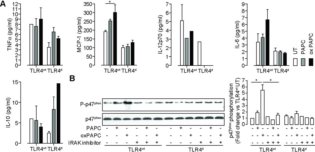
Figure 8.
TLR4 triggers inflammatory cytokine release and promotes IRAK modulated p47phox phosphorylation in response to oxidized phospholipid treatment in BMDM derived from TLR4wt and TLR4d mice. (A) BMDM were treated with PAPC and oxPAPC and inflammatory cytokine levels in the supernatant were determined. (n=3/group; *p<0.05) (B) These blots show p47phox expression and phosphorylation in response to PAPC and oxidized PAPC treatment. Lysates from bone marrow derived monocytes isolated from TLR4wt and TLR4d mice were immunoblotted for p47phox and phospho-p47phox (left). A subset of experiments was performed in presence of an IRAK inhibitor. The figure on the right represents the photodensitometric quantification of the blots. (n=3) Data are mean ± SD. (*p<0.05).

