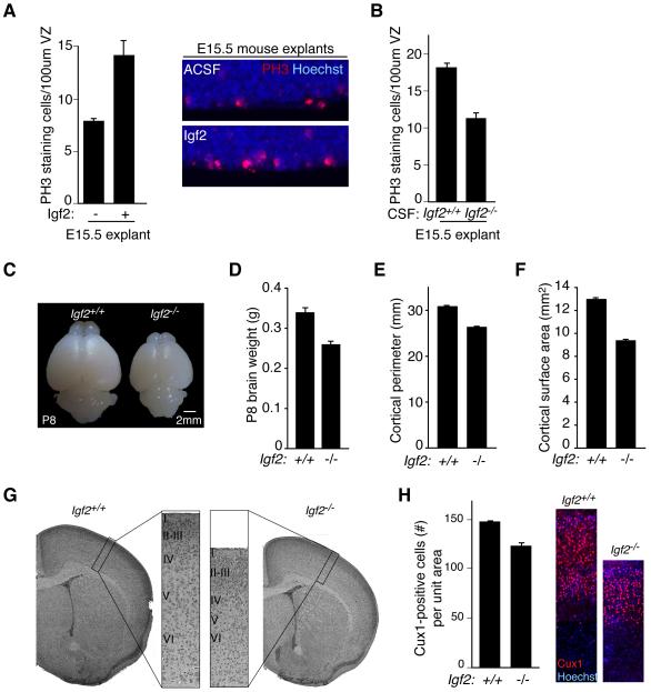
Figure 5. CSF Igf2 regulates progenitor proliferation and brain size.
(A) Left panels: E15.5 C57BL/6 explants cultured in NBM supplemented with 20% ACSF or ACSF/Igf2. Igf2 stimulated the proliferation of PH3-positive cortical progenitor cells (C57BL/6 explants: ACSF mean: 7.4 ± 0.2; E16.5 mean: 14.1 ± 1.4; Igf2 mean: 11.2 ± 0.3; Kruskal-Wallis; E16.5 vs. ACSF, p<0.01; Igf2 vs. ACSF, p<0.05; E16.5 vs. Igf2, N.S.; n=3). Right panels: Representative images of explants quantified in left panels. (B) E15.5 C57BL/6 explants cultured in NBM supplemented with 20% ACSF or E16.5 Igf2−/− CSF. Igf2-deficient CSF failed to stimulate progenitor cell proliferation compared to control (ACSF: 17.9 ± 0.8; Igf2−/− CSF: 11.4 ± 1.0; Mann-Whitney; p<0.06; n=3 and n=4, respectively). (C) Representative images of P8 Igf2−/− and control brains. (D) Igf2-deficiency reduced P8 brain weight (Igf2+/+: 0.34g ± 0.008; Igf2−/−: 0.26g ± 0.004; Mann-Whitney, p<0.0001, n=11). (E) Igf2-deficiency reduced P8 cortical perimeter (Igf2+/+: 30.9mm ± 0.01; Igf2−/−: 26.4mm ± 0.1; Mann-Whitney, p<0.0001, n=11). (F) Igf2-deficiency reduced P8 cortical surface area (Igf2+/+: 13.0mm2 ± 0.1; Igf2−/−: 9.4mm2 ± 0.1; Mann-Whitney, p<0.0001, n=11). (G) H&E staining of Igf2−/− and control brains at P8. (H) Left panels: Igf2−/− brains have reduced numbers of upper layer neurons marked by Cux1 (Total Cux1-positive staining cells in equally sized cortical columns expressed as mean ± S.E.M.: Igf2+/+: 157 ± 1.5; Igf2−/−: 131.3 ± 3.3; t-test, p<0.005, n=3). Right panels: Representative images of Igf2−/− and control brains quantified in left panels. See also Figure S3.

