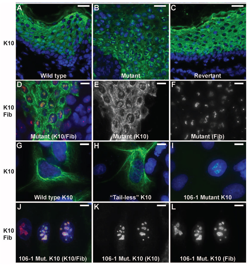
Fig. 4.
Mutant keratin 10 is redirected to nucleoli in vivo and in vitro. (A–C) Images of normal, mutant and revertant skin stained with DAPI and antibodies to keratin 10 reveal discrete foci of nuclear keratin 10 in mutant skin which are absent in normal and revertant skin. (Scale bars = 50 µm). (D–F) Co-staining with the nucleolar marker fibrillarin shows that keratin 10 is in the nucleolus. (Scale bars = 10 µm) (G–I) Constructs bearing wild-type keratin 10 (G), keratin 10 truncated at the beginning of the tail domain (amino acid 459) (H) and keratin 10 with the kindred 106 frameshift mutation beginning at codon 460 (I) were expressed in PLC cells and stained with DAPI and monoclonal antibody to keratin 10. Wild-type and ‘tailless’ K10 integrate into the cytoplasmic filament network while the IWC mutant K10 localizes to nucleoli as shown by costaining with fibrillarin (J–L). (Scale bars in G–L = 10 µm).

