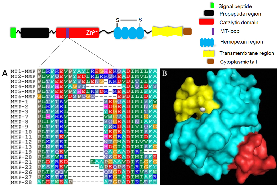
Fig. 1.
(A) Partial sequences alignment for MMPs. Numbering is according to the sequence of MT1-MMP. The peptide sequence 160REVPYAYIREGHEKQ174 is unique to MT1-MMP. Amino acids are represented as single-letter codes. The gap (represented by “−”) is auto-generated by the BioEdit software. (B) The 3D structure of MT1-MMP is shown as an electrostatic potential surface with the target peptide sequence 160REVPYAYIREGHEKQ174 colored in red. The catalytic center, in yellow, is located on the surface of the MT1-MMP. Zinc is shown in white.

