Figure 5.
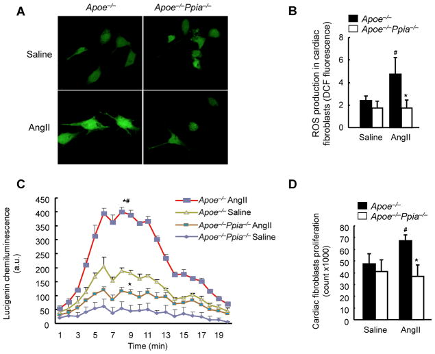
CyPA activates cardiac fibroblasts by enhancing ROS production. (A) Representative DCF staining of mouse cardiac fibroblasts. AngII-induced ROS generation after 4 hours was decreased in CyPA-deficient cardiac fibroblasts. (B) Densitometric analysis of DCF fluorescence in response to AngII shows significant reduction in Ppia−/− cardiac fibroblasts at 4 hours (n = 8 in each group). (C) Superoxide production in cardiac fibroblasts exposed to lucingenin for 4 hours. Results are mean ± SD of three independent experiments performed in triplicate. # equals P< 0.05 in Saline versus AngII; *equals P< 0.05 in Apoe−/− versus Apoe−/− Ppia−/− mice. (D) Proliferation of cardiac fibroblasts. Apoe−/− and Apoe−/− Ppia−/− fibroblasts were treated with saline or AngII. After 48 hours of incubation, cells were counted (n = 3 in each group). Results are mean ± SD. # equals P< 0.05 in Saline versus AngII; *equals P< 0.05 in Apoe−/− versus Apoe−/− Ppia−/− cardiac fibroblasts.

