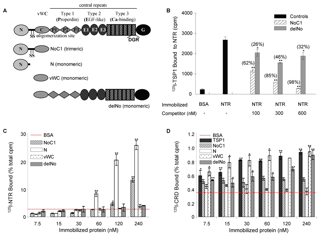
Figure 3. Localization of the sFRP-1 binding site in TSP1.
20 µg/ml of recombinant NTR domain were absorbed onto microtiter plate wells (B), and 125I-TSP1 binding was measured at 37°C in the presence of competing concentrations of the indicated unlabelled recombinant regions of TSP1 (as shown in A). Nonspecific sites were blocked with 3% (w/v) BSA in DPBS containing Ca2+ and Mg2+. Data were normalized and presented in brackets as percent of inhibition. The results are representative of three independent experiments, each performed in triplicate. 125I-NTR domain (C) and 125I-CRD (D) binding to the indicated concentrations of TSP1 or TSP1 recombinant regions coated onto microtiter wells was determined as above. Data were normalized and are presented as percent of total cpm. The results are representative of four (125I-NTR) and two (125I-CRD) independent experiments, each performed in triplicate.

