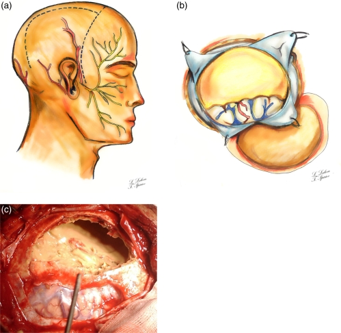
Figure 2.
a. Schema of skin incisions and related superficial neuro-vascular anatomy. A large right fronto-temporo-parital craniotomy was made; b. Schema of the hematoma wall, after dural incision. The haematoma had clear margins and was separated from the surrounding brain (right temporal lobe is visualized); c. Intraoperative image after craniotomy and dural incision. The parietal hematoma wall was excised and the yellowish liquefied content is seen. The haematoma had clear margins and was separated from the surrounding brain

