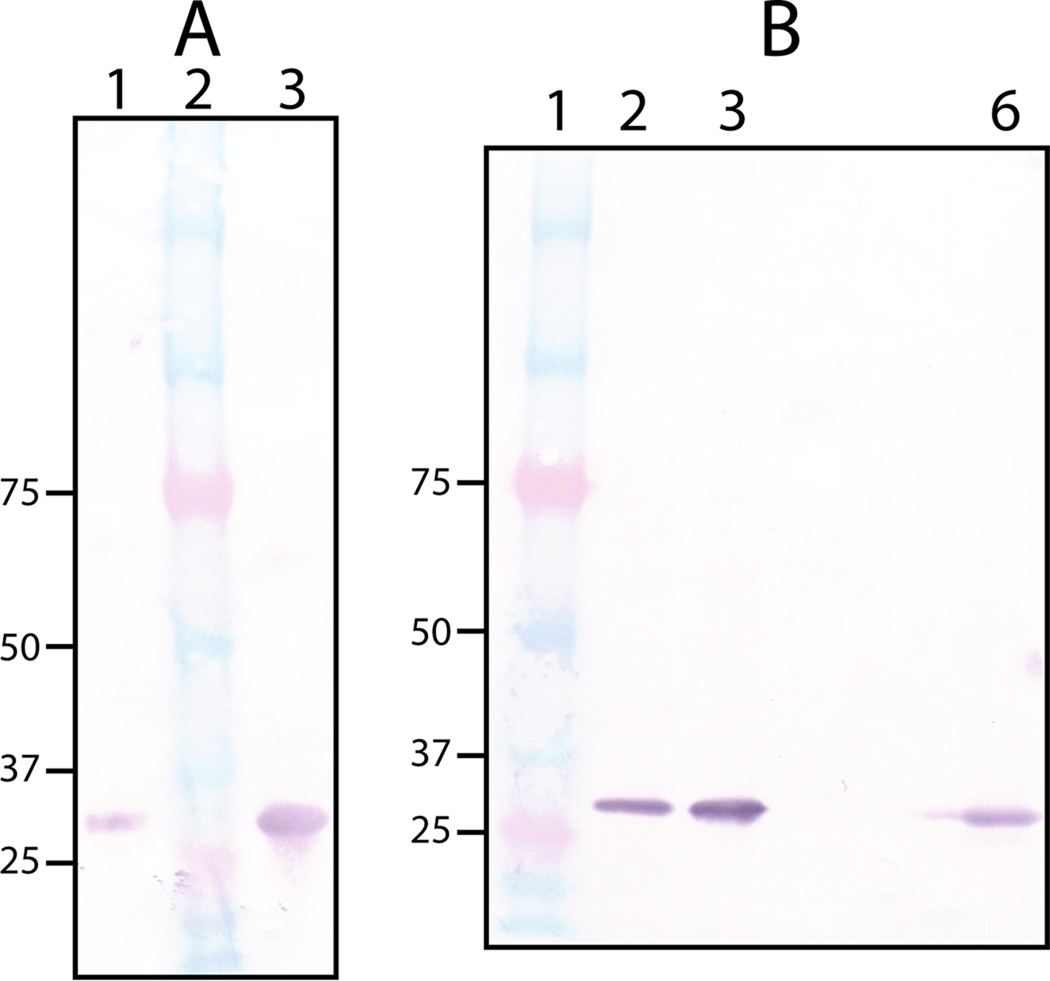
Fig. 5.
Western blot analysis using anti-rPmCCAP IgG. (A) Detection of purified rPmCCAP (lane 1) and rP4 (lane 3) using a 1:80,000 antiserum dilution. Lane 2 contains molecular weight standards. (B) Analysis of clinical P. multocida and M. haemolytica clinical strains. Lane 1 contains molecular weight standards. Lanes 2 and 3 contain cell pellet fractions of P. multocida clinical strains cultured from avian and porcine sources, respectively. Lane 6 contains the cell pellet fraction from a M. haemolytica clinical strain (bovine source). The antiserum was diluted 1:160,000 in each case.

