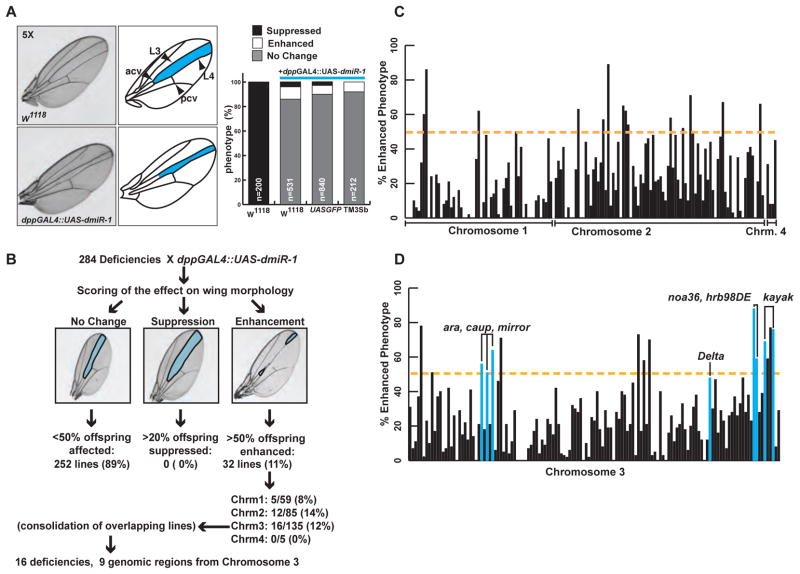
Figure 1. Misexpression of dmiR-1 in the Drosophila wing results in a distinctive phenotype and identifies genetic partners of dmiR-1.
(A) Upper left: image of a wildtype wing, with pertinent anatomical structures labeled to the right. L3: long vein number 3, L4: long vein number 4, acv: anterior cross vein, pcv: posterior cross vein. Area highlighted in blue: area of expression of the wing disc specific decapentaplegic (dpp) enhancer. Lower left: image of wing from a dpp-GAL4::UAS-dmiR-1 mutant fly. Note narrowing of the L3/L4 inter-vein distance. Left: Incidence of enhancement, and suppression of the wing phenotype seen in dpp-GAL4::UAS-dmiR-1 mutant flies when mated with control fly lines. W1118: wildtype. The lines UAS-GFP and dpp-GAL4::UAS-dmiR-1/TM3Sb (TM3Sb) served as controls. TM3Sb: balancer.
(B) Summary of results from the forward genetic screen.
(C) Distribution of percentage affected offspring from each deficiency, organized by chromosome (1, 2, and 4). Yellow dashed line: point at which 50% of progeny were affected. Those deficiencies that met or exceeded this level were considered strongly positive (p<0.0001).
(D) Distribution of percentage affected offspring from each deficiency, organized by position on chromosome 3, going left to right. Blue bars: deficiencies containing specific genes (labeled) that enhanced the wing phenotype.
(See also Supplementary Table 1)

