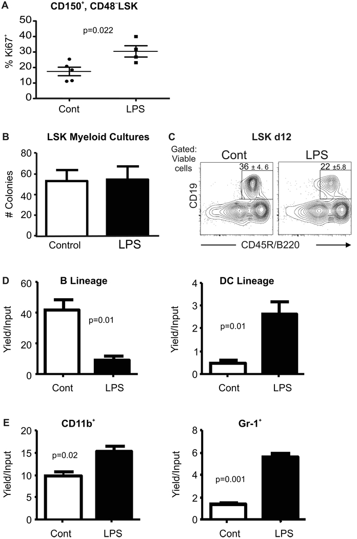
Figure 2. Primitive hematopoietic cells are altered by chronic LPS exposure.
A, Cell cycling activity in CD150+ CD48− LSK in control and LPS treated marrow was assessed by flow cytometry for the Ki-67 proliferation antigen. B, LSK were sorted from mice at the end of LPS or PBS exposure and placed in myeloid supporting Methocel cultures. Average numbers of colonies were counted 10 days later. C, The same fractions were placed in defined, lymphoid supporting liquid cultures for 12 days before flow cytometry. Boxes show percentages of B220+CD19+ lymphocytes that were generated; (p=0.02). This data is representative of that found in four independent experiments with 5–8 mice per group. All error values depict SEM. D, Common lymphoid progenitors (CLP) defined as in Supplemental Fig. 1B were recovered from the two groups of mice after 4–6 weeks of chronic exposure and sorted to high purity. These were placed in defined lymphoid supporting cultures (see Experimental Procedures) for 8 days before harvest and flow cytometry. Yields of CD19+B220+ B-lineage lymphocytes and CD11c+ dendritic cells ±SEM per input progenitor were calculated. E, Common myeloid progenitors (CMP) sorted as shown in Supplemental Fig. 1B were placed in myeloid supporting cultures and their potential for generation of myeloid marker bearing cells was determined 8 days later by flow cytometry. Absolute yields of myeloid cells ±SEM per input progenitor are shown. Similar results were obtained in four independent experiments with at least four culture wells per group. Statistical significance was determined by t-test analysis.

