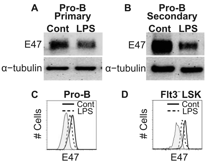
Figure 4. Chronic TLR stimulation leads to decreased E2A protein abundance in pro-B and primitive cells.
Western blot analysis comparing E47 protein levels in pro-B cells generated from control or LPS treated marrow after primary (A) and secondary (B) transplants. α-tubulin served as an internal protein control for this assay. The data is representative of three independent experiments per time point. Two independent flow cytometry analyses from separate laboratories assessed intracellular E47 protein levels in individual pro-B (C) and HSC-enriched Flt3−LSK cells (D) after secondary transplantation. Plots are representative of 3 independent experiments with 5–6 mice per analysis. Statistical significance was determined by t-test analysis.

