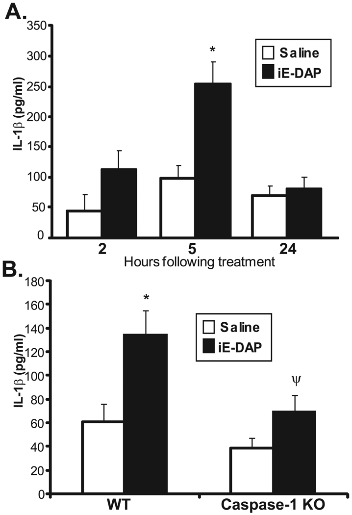
FIGURE 5.
Activation of NOD1 by iE-DAP results in increased IL-1β in the mouse eye by caspase-1. Mice were treated with intravitreal injection of saline or 100 µg iE-DAP. Cytokine production of IL-1β in eye tissue homogenates was measured by ELISA as a function of time after treatment (A) or in caspase-1 knockout (KO) mice (B) 5 hours after treatment. Data are mean ± SEM (n = 6 mice/treatment/time/genotype). *P < 0.05 comparison between saline and iE-DAP treatments within a genotype. ψP < 0.05 (comparison between iE-DAP–treated wild-type and caspase-1 KO mice).

